
Polarization-Based Optical Imaging for Clinical Diagnostics
Edited by Yurii Ushenko Edited by Weidong Dou Edited by Oleksandr Ushenko Edited by Oleksandr Dubolazov Edited by Iryna Soltys
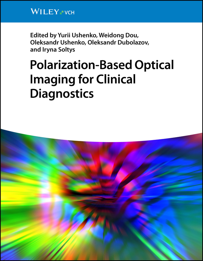
Hardback (Forthcoming)
(October 2026)
ISBN: 9783527414697
8.5 x 10.91 inches
Price: $212.00
On back order
“Polarization-Based Optical Imaging for Clinical Diagnostics” presents a comprehensive methodology for optical mapping of biological tissues and fluids using 3D Jones-Mueller digital holography. It covers physical principles, experimental systems, and data analysis algorithms, offering a unified theory for polarization-based diagnostics. The book includes applications in oncology, cardiology, gynecology, and forensic medicine, demonstrating its clinical relevance. What sets this book apart is its integration of statistical, fractal, and wavelet analyses with advanced imaging techniques, enabling precise differentiation of pathological states. It also features software tools and image libraries, making it both a reference and a practical guide for modern biomedical diagnostics.
“Polarization-Based Optical Imaging for Clinical Diagnostics” presents a comprehensive methodology for optical mapping of biological tissues and fluids using 3D Jones-Mueller digital holography. It covers physical principles, experimental systems, and data analysis algorithms, offering a unified theory for polarization-based diagnostics. The book includes applications in oncology, cardiology, gynecology, and forensic medicine, demonstrating its clinical relevance. What sets this book apart is its integration of statistical, fractal, and wavelet analyses with advanced imaging techniques, enabling precise differentiation of pathological states. It also features software tools and image libraries, making it both a reference and a practical guide for modern biomedical diagnostics.
PART I: METHODS AND SYSTEMS OF OPTICAL MAPPING IN BIOMEDICAL RESEARCH
1. Overview of modern optical mapping techniques
1.1 Diffuse, fluorescence molecular, bioluminescence, photoacoustic, laser coherent, andlaser polarization-sensitive systems
1.2 Unsolved problems and proposed solutions via 3D Jones-Mueller digital holographic mapping
PART II: PHYSICAL PRINCIPLES, ALGORITHMS AND METHODS OF 3D JONES-MUELLER DIGITAL HOLOGRAPHIC MAPPING
2. Physical principles of 3D Jones-Mueller digital holographic mapping
2.1 Phenomenological models of optically anisotropic tissue and fluid samples
2.2 Reconstruction of optical anisotropy parameters (linear and circular birefringence and
dichroism)
3. Algorithms and methods of 3D Jones-Mueller digital holographic mapping
3.1 Digital holographic reconstruction and phase scanning
3.2 Multichannel polarization-interference measurement and reconstruction of anisotropy
maps
4. Basic algorithms for processing 3D Jones-Mueller digital holographic mapping data
4.1 Statistical analysis
4.2 Correlation analysis
4.3 Wavelet analysis
4.4 Multifractal analysis
4.5 Singular analysis
PART III: BIOMEDICAL AND CLINICAL APPLICATIONS OF 3D JONES-MUELLER DIGITAL HOLOGRAPHIC MAPPING
5. Diagnostic efficiency of 3D Jones-Mueller digital holographic mapping
5.1 Phase waves of local depolarization in biological tissues
5.2 Differential Mueller-matrix mapping of polycrystalline tissue components
5.3 Optical interference approach for tumor differentiation
5.4 Wavelet-enhanced polarimetry for cancer analysis
5.5 Holographic scanning of myocardial layers
5.6 Holographic histology for necrotic tissue differentiation (forensics)
5.7 Imaging of blood film microstructure
6. Diagnostic efficiency of 3D Jones-digital holographic mapping
6.1 Mapping of biological fluid facies
6.2. Jones matrix tomography
6.2 Multifractal scanning for COVID-19 diagnostics
6.3 Scanning of blood plasma films for organ pathology
6.4 Polarimetry of blood films for thyroid pathology
Appendices
A1. Programs for 3D Jones-Mueller holographic reconstruction
A2. Programs for layer-by-layer reconstruction of polycrystalline structures
A3. Programs for statistical and correlation data analysis
A4. Album of Mueller matrix images
A5. Album of optical anisotropy maps
Conclusion
- Edited by Yurii Ushenko
- Edited by Weidong Dou
- Edited by Oleksandr Ushenko
- Edited by Oleksandr Dubolazov
- Edited by Iryna Soltys
Similar Books



 Recycling and Regeneration of Spent Lithium-Ion Batteries
Technologies, Applications, and Sustainable Futures


 Medicinal Chemistry of Plant-Derived Natural Products
A Chemical Exploration of Plants as a Source of Modern Drugs


 Ernst Haeckel: Ausgewählte Briefwechsel. Historisch-kritische Ausgabe. Bd. 8
Briefwechsel mit Carl Gegenbaur. Wissenschaftskorrespondenz. Mai 1858 – Juli 1902


